

 Sequencer Products: SEQ ALL


 Technologies
 Applications
 Online Resources
 Data Bulletins
 Service & Support
 Global Programs
 Introduction
 Newsroom
 Doing Business With Us
 Creative Club


The "Product FAQs" column collects products related questions which MGI customers inquiries frequently. And we organizes FAQs which will be released in the future to help you understand and use MGI products better.
Q1.What features have been added to the DNBSEQ-G400RS FluoXpert upgrade?
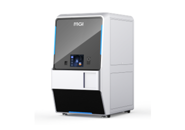
DNBSEQ-G400RS FluoXpert is a multifunctional machine that integrates high-throughput sequencing technology (MPS) and multiplex fluorescence technology (mIF). As both functions were implemented based on the same set of hardware, it can switch between gene sequencing and multiplex immunofluorescence staining simply by opening different programs.
This can become complementary to the sequencing technology, enabling a full range of analyses from the protein level and gene level. The combination of both technologies on a single machine can provide a more accurate and comprehensive evaluation basis for the tumor immune microenvironment.
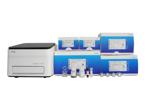
Q2. What is the product composition of DNBSEQ-G400RS FluoXpert?
DNBSEQ-G400RS FluoXpert includes the full sets of hardware equipment from DNBSEQ-G400RS, FluoXpert mIF set, FluoXpert program and FluoXpert Vision image analysis platform.
Q3. What are the advantages of the multiplex immunofluorescence function of DNBSEQ-G400RS FluoXpert?
DNBSEQ-G400RS FluoXpert has high compatibility. It is compatible with both frozen tissue and FFPE samples. It is also compatible with commercially available antibodies without complex conjugation process. Both staining and imaging processes are all automatically completed, enabling multiple biological markers to be detected on a tissue section while retaining spatial information.
With FluoXpert mIF sets, DNBSEQ-G400RS FluoXpert supports the detection of 3-24 markers on the same tissue section, which can be widely used in tumour typing, prognosis evaluation, tumor microenvironment research, neuroscience, translational medicine research, etc.
Based on the design concept of staining and imaging integration, systematization, and modularization, the preprocess of DNBSEQ-G400RS FluoXpert is fast and convenient. Moreover, an autofluorescence removal reagent is integrated into the FluoXpert mIF set, which greatly simplifies the sample processing steps before loading.
DNBSEQ-G400RS FluoXpert contains a highly stable staining and imaging process which showed no significant difference from the gold standard IHC staining effect. Also, the optimized stitching algorithm and imaging technology can achieve subcellular level resolution panoramic imaging effects.
Q4. What are the advantages of the gene sequencing function of DNBSEQ-G400RS FluoXpert?
The multiplex staining function of DNBSEQ-G400RS FluoXpert does not affect the sequencing function but complements each other. Compared with the regular protein staining and imaging machine, the sequencing data of DNBSEQ-G400RS FluoXpert can provide one-step sequencing data as a supplement or reference for the staining results.
Compared with regular genome sequencers, DNBSEQ-G400RS FluoXpert has superior sequencing performance, supports multiple sequencing read lengths, flexible throughput (55Gb-1440Gb), supports different specifications of sequencing carriers to run independently, can fully meet a wide range of sequencing needs, and is one of the preferred models for sequencing laboratories around the globe.
Q5. How to upgrade to FluoXpert for those who have purchased DNBSEQ-G400RS?
Users who have purchased DNBSEQ-G400RS only need to order the “Multi-Omics Package”, without the need to buy a new machine. The composition of the multi-omics package includes: FluoXpert program and FluoXpert Vision image analysis platform (optional).
The FluoXpert mIF set can be purchased as needed according to the experiment. At present, two versions of these reagent sets are provided: standard version (6-plex, 24-plex) and customized version (depending on project needs, can be selected within the range of 3-24 plex).
The FluoXpert mIF set (standard version) includes: a FluoXpert mIF reagent kit, and 5 FluoXpert staining flow cells. Considering the staining needs of different size sections, DNBSEQ-G400RS FluoXpert provides three optional slide specifications, including:
-SS (Solo Small, suitable for small sections): Suitable for small tissue sections, such as puncture small tissues. Faster in speed.
-SL (Solo Large, suitable for large sections): Suitable for large tissue sections, such as tonsils and large tumors. Larger area and field of view.
-DL (Dual Large, suitable for double section co-staining): One slide can hold two sections, suitable for batch staining. Saves reagents and time.
Q6. How are the number of staining cycles and the best throughput of DNBSEQ-G400RS FluoXpert calculated?
Take the standard version reagent set (6-plex) as an example. To complete the 6-plex staining run, DNBSEQ-G400RS FluoXpert needs to perform 3 cycles of antibody staining and 1 cycle of nuclear staining (2 markers for each single staining cycle). This adds up to a total of 4 staining cycles with the total time being about 4 hours. The calculation of the best throughput is based on a 5 days a week and 8 hours per day meaning that the staining runs can be performed 2 runs a day and 2 sections per run (SS/SL), adding to 2*2*5=20 sections(SS/SL slides), or 4*2*5 = 40 sections (DL slides) completed per week of work time.
Q7. What is the result format generated from FluoXpert?
The data generated by FluoXpert is a stitched whole-tissue image. This image will crop continuous tissue areas (optional) and output different compression formats (OME-TIFF, TIFF, JPEG, PNG, BMP).
Q8. What kind of analysis results can the FluoXpert Vision platform provide?
The FluoXpert Vision image analysis platform can provide image visualization and annotation, perform positive cell count quantitative analysis based on thresholds, perform cell phenotype analysis based on the co-expression of multiple markers, and perform spatial location analysis of adjacent cells.
Q9. How to switch between sequencing and immunofluorescence modes?
Currently, the instrument cannot support the opening sequencing and immunofluorescence software at the same time. One needs to be completely closed before the other start.
Q10. What is the effective scanning area of the staining carrier?
The scanning area of the SS staining flow cell is 15mm*15mm, the SL staining flow cell is 15 mm* 20 mm, and the DL staining flow cell is 15 mm*20 mm*2.