About MGI
MGI Tech Co., Ltd. (or its subsidiaries, together referred to MGI), is committed to building core tools and technologies that drive innovation in life science. Our focus lies in research & development, manufacturing, and sales of instruments, reagents, and related products in the field of life science and biotechnology. We provide real-time, multi-omics, and full spectrum of digital equipment and systems for precision medicine, agriculture, healthcare and various other industries.

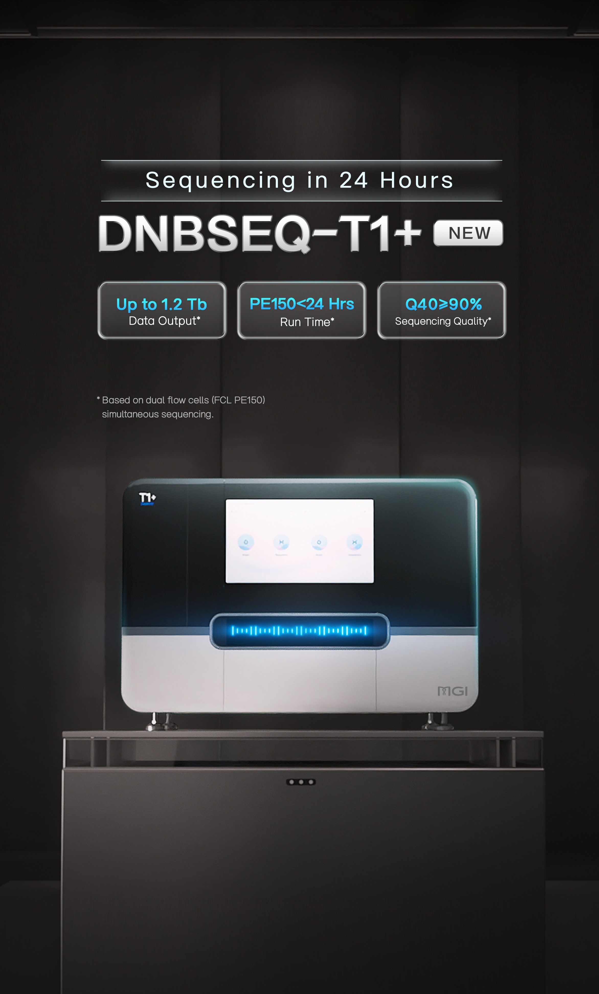
 Sequencer Products: SEQ ALL
 Technologies
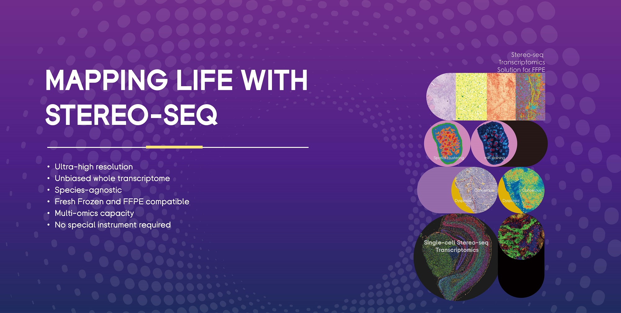
 Applications
 Online Resources
 Data Bulletins
 Service & Support
 Introduction
 Newsroom
 Doing Business With Us
 Creative Club