

 Sequencer Products: SEQ ALL


 Technologies
 Applications
 Online Resources
 Data Bulletins
 Service & Support
 Global Programs
 Introduction
 Newsroom
 Doing Business With Us
 Creative Club


The "Product FAQs" column collects products related questions which MGI customers inquiries frequently. And we organizes FAQs which will be released in the future to help you understand and use MGI products better.
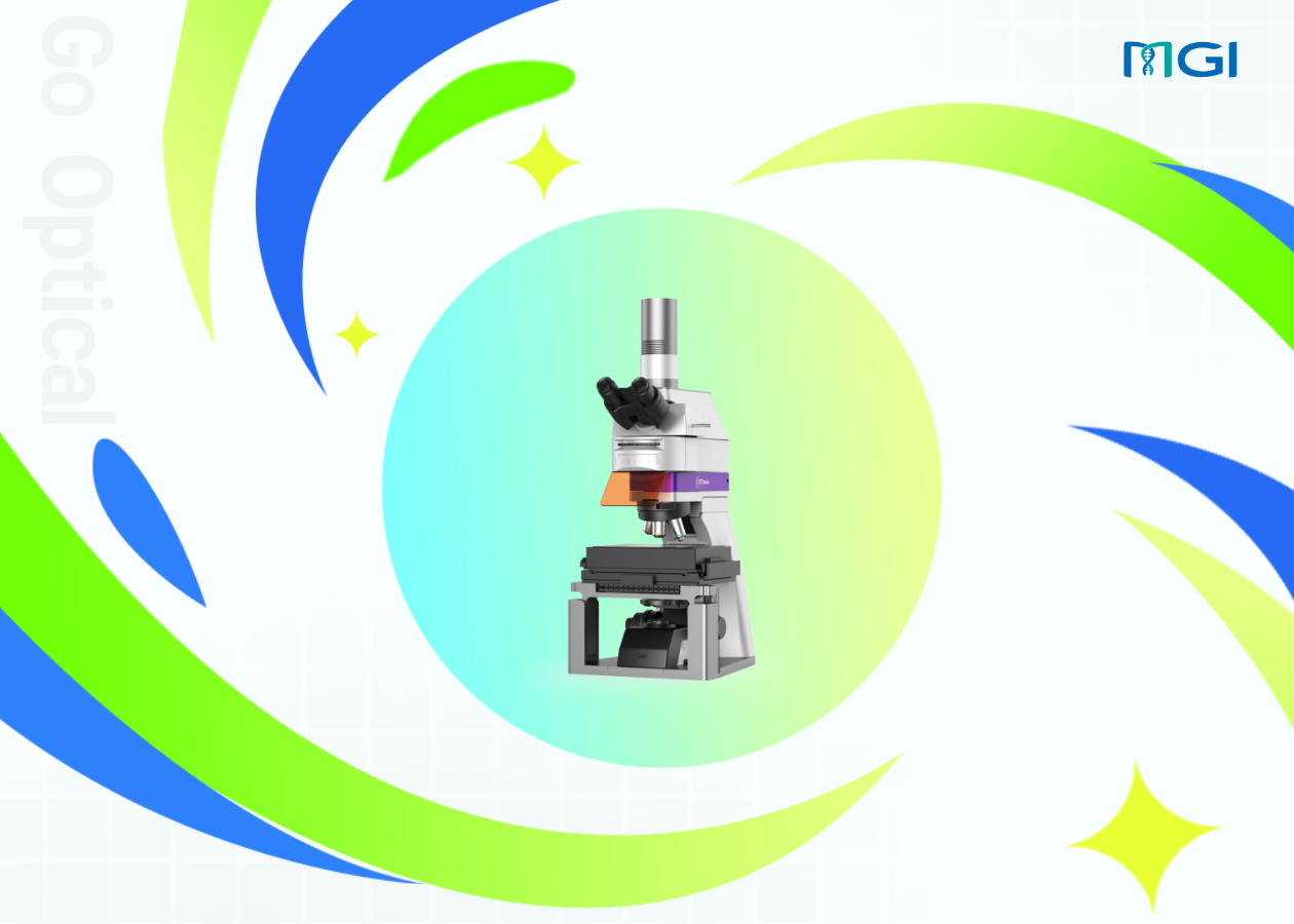
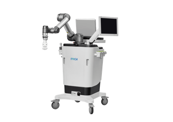
Q1:What is the STOmics Microscope Go Optical?
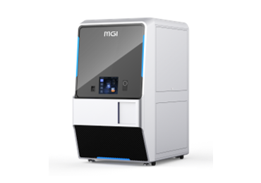
The STOmics Microscope Go Optical is a precision instrument that integrates multidisciplinary technologies such as optics, mechanics, electronics, and computer science. It operates by controlling the microscopic imaging system and sections on slides or chips to move in a defined pattern, capturing multiple continuous high-resolution microscopic images, which are then seamlessly spliced together to generate a high-resolution whole-slide image. Once the traditional sections are transformed into a high-resolution digital image, users can view the sections anytime, anywhere on a computer or mobile device without the need for a microscope. This digital image offers several advantages, including non-fading, ease of preservation, management, sharing, and the ability to zoom in and out for a comprehensive view. It has a wide range of applications in fields such as pathological diagnosis, educational training, drug research, and scientific research.
Q2:What are the advantages of the STOmics Microscope Go Optical?
1.Short scanning time: When scanning the 10mm * 10mm area under the 10X objective lens, the scanning time of the microscope shall not exceed 70 seconds. This reduced scanning time ensures the integrity of the sample's RNA is maximally preserved.
2.Large travel range: The Go Optical is compatible with scanning of mainstream sample sizes of 10mm by 10mm and 50mm by 30mm, with the capability to scan samples up to 100mm by 70mm. This meets the scanning requirements for the majority of large STOmics chips.
3. High stitching Precision: With a 10X objective lens, the Go Optical achieves a stitching precision of 5 μm. This means that when multiple images are stitched together, the alignment between the images will be highly accurate, enhancing the precision and efficiency of observation.
Q3:What are the main components of the STOmics Microscope Go Optical?
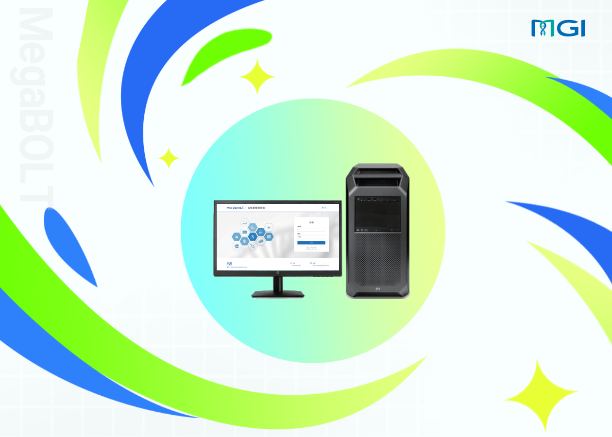
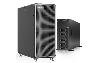
Go Optical including 2 parts, hardware and software. The hardware comprises a microscope, an electronic control box, a computer host, and a display monitor. 2 Software comprises Scanner (for image scanning) and Tiff Browser (for image viewing).
Q4:What are the main applications of the Go Optical?
The Go Optical is primarily recommended for scanning and imaging of chips/slides in various application scenarios of spatiotemporal omics research.
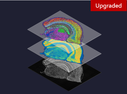
Q5:What are the main application scenarios in spatiotemporal omics?
The Go Optical is primarily designed for various application scenarios within spatiotemporal omics, including:
1)Fluorescence imaging mode for capturing fluorescence-labeled silicon chip samples;
2)Brightfield imaging mode for capturing pathology-stained silicon chip samples;
3)Transmission brightfield imaging mode for capturing pathology-stained glass slide sample.
Q6:How to choose Go Optical?
Go Optical has two Cat. No.. The Cat. No. of 900-000586-00 is the RUO-China and RUO-CE version; The Cat. No. of RUO-NRTL is RUO-NRTL version.
Q7:Are there any optional accessories? How should they be selected for purchase?
The standard configuration of the Go Optical includes objective lenses of 4X, 10X, and 20X only. Customers have the option to select a 40X objective lens to meet their specific requirements, with the Cat. No. of 060-000217-00.
The imaging principle of the Go Optical is similar to that of a traditional fluorescence microscope. The microscope uses a point light source with high luminous efficiency, which, after passing through the illumination system and filter block, emits light of a certain wavelength as excitation light. This excitation light stimulates fluorescent substances within the tissue sample to emit fluorescence. The fluorescence is then magnified through the objective lens and tube lens (or eyepiece) for imaging (or direct observation by the human eye) on a camera.
Q9:What are the illumination sources for the Go Optical? What observation methods does it support?
There are two illumination sources, including epi-illumination solid-state continuous white light source and transmitted light source. Go Optical supports various observation methods such as epifluorescence, epi-brightfield, and trans-brightfield, which can be selected based on different sample types and observation requirements.
Q10:What types of fluorescent dyes does Go Optical support?
The standard types of fluorescent dyes used in the Go Optical included DAPI, FITC, TRITC, and CY 5. In addition, other dye filter blocks are supported to meet the wider range of fluorescence observation requirements.
Q11:What are the optional modes for automatic focus detection?
The automatic focus detection function provides two optional modes: Auto Focus Mode and Map Navigation Mode. The auto focus mode automatically finds the best focus through the photoelectric sensor, while the map navigation mode allows the user to manually select a specific observation area for focus detection.
Q12:What are the technical advantages of the Go Optical compared to other brands of scanning fluorescence microscope in the application of spatiotemporal omics?
The imaging is clear with minimal stitching errors; automatic focus detection for rapid imaging; seamless stitching with a high-precision algorithm for eliminating stitch shadows, creating a panoramic microscopic image without visible seams.
Q13:What is the operational requirement of Go Optical?
For indoor use, the Go Optical must be placed on a stable and reliable table, or worked on an anti-vibration workbench. The temperature requirement is 5℃ to 40℃, and the relative humidity requirement is 30%RH to 75%RH, with no condensation. For detailed requirements, please contact the after-sales engineer for provision.